ASCP-MLT ASCP MEDICAL LABORATORY TECHNICIAN - MLT(ASCP) Free Practice Exam Questions (2026 Updated)
Prepare effectively for your ASCP ASCP-MLT MEDICAL LABORATORY TECHNICIAN - MLT(ASCP) certification with our extensive collection of free, high-quality practice questions. Each question is designed to mirror the actual exam format and objectives, complete with comprehensive answers and detailed explanations. Our materials are regularly updated for 2026, ensuring you have the most current resources to build confidence and succeed on your first attempt.
Unexplained bleeding is associated with immediate hemolytic transfusion reactions, but is not usually associated with delayed hemolytic transfusion reactions. The bleeding results from disseminated intravascular coagulation (DIC) due to ABO antibodies causing intraventricular hemorrhage (IVH).
Which of the following signs and symptoms may be associated with immediate transfusion reaction, but is NOT usually associated with delayed hemolytic transfusion reaction?
Streptococcus pyogenes is the correct answer. Streptococcus pneumoniae is alpha-hemolytic which demonstrate a green area of partial hemolysis around the colonies. Staphylococcus aureus will produce complete hemolysis but is catalase positive. Streptococcus agalactiae is beta-hemolytic but only produces a partial clearing of the red blood cells in sheep blood agar.
Complete hemolysis of sheep blood agar as demonstrated by the image below would be seen in which of the following catalase-negative isolates?

The risk of HbS polymerization is enhanced by a low (acid) pH, a state of dehydration, and increased levels of 2,3-DPG. Increased temperature (above 37°C) also adds to the risk.
Hematology
Which group of conditions INCREASES the risk of HbS polymerization?
HLA-DR is a class II MHC.
HLA-A, HLA-B, and HLA-C are all class I MHC.
Which of the following antigens is classified as a Major Histocompatibility Complex
Class II antigen (MHCII)?
This is the lens power of the scanning objective lens.
The correct answer which best fits the characteristics in this question is Mycobacterium tuberculosis.
What is the MOST likely identity of an acid-fast bacilli that showed buff colored, dry, heaped colonies; niacin, nitrate, and urease positive; and took 23 days at 35ºC to grow?
Angiotensin is an oligopeptide in the blood that causes vasoconstriction, increased blood pressure, and release of aldosterone from the adrenals.
A major action of angiotensin II is:
A cardiac risk profile is performed in:
This drawing depicts beta thalassemia minor B+/B. In Beta thalassemia minor B+/B, one beta gene locus is partially deleted or inactive.
Hematology
This drawing depicts which beta chain genotype ?
Neutrophils, lymphocytes and macrophage/ monocytes can be found in all types of body fluid differentials. Bronchial cells can be found only in bronchial washings and BAL specimens. Mesothelial cells are found only in serous body fluids including pleural fluid, peritoneal fluid, and pericardial fluid.
Select the specific cells listed below that can be found in all types of body fluid.
The two main areas of the clinical laboratory are:
HbsAg is positive in acute and chronic Hepatitis B infections, since the antigen is found on the actual surface of the virus. HbeAg is present in the blood when the hepatitis B viruses are replicating, indicating an active infection. Anti-Hbc IgM is present due to the immune response to the presence of the hepatitis core antigen and indicates an acute infection. Anti-HBs is generally interpreted as indicating recovery and immunity from hepatitis B virus infection, according to the CDC.
Given the following results, what is the immune status of the patient?
HbsAg: positive
HbeAg: positive
Anti-HBc IgM: positive
Anti-HBs: negative
The primary mechanism responsible for glomerular filtration is:
Rule-out is a process by which antibodies are identified as being unlikely in a given sample because of the absence of an expected antigen-antibody reaction. In other words, the absence of a reaction is noted with a cell that is positive for the corresponding antigen.
Although rule-out procedures may vary somewhat from institution to institution, the following general principles apply:
Non-reactive cells are selected for rule-out. To be classified as non-reactive, a cell must NOT have reacted at any phase of testing in a given panel or screen.
Using the logic that if the rule-out cell is positive for a given antigen, it should have reacted with the corresponding antibody, you can rule-out antibodies that correspond to antigen positive cells.
To increase the probability that rule-out will not mistakenly eliminate a weakly-reacting antibody that exhibits dosage*, use only cells that are homozygous for the corresponding antigen for those systems that generally show dosage. Generally these include: C, c, E, e, Fya, Fyb, Jka, Jkb, M, N, S, and s.
In this case, it is only possible to rule out on screening cell 2 since it demonstrates a negative reaction with the patient serum. Anti-C cannot be ruled out since the C antigen is heterozygous on screening cell 2 with c. Anti-Fya cannot be ruled out since this antigen is not present on screening cell 2. Anti-M and anti-Jka can be ruled out since the antigens are homozyous while demonstrating a negative reaction on screening cell 2.
Rule-out, while very useful, can lead to error. Ruling out an antibody should be combined with other supporting data to increase confidence in the solution; the more data collected, the higher the probability that the final solution is correct.
*Dosage means that there are two "doses" of the same antigen present on the red cells . Antibodies that exhibit dosage react more strongly with homozygous cells (e.g., Jka Jka) than with heterozygous cells (e.g., Jka Jkb) .
Based on the phenotype of the RBC screening cells, and patient results shown on the right, which of the following antibodies CANNOT be ruled out?
Primary- Target glands (such as thymus, thyroid, parathyroid, etc.)
Secondary- Pituitary gland
Tertiary- Hypothalamus
Match the type of endocrine dysfunction with the appropriate organ:
1. Target gland
2. Pituitary gland
3. Hypothalamus
Which of the following is NOT part of the stand or framework of the microscope?
Oxalate, EDTA, and citrate are anticoagulants that inhibit clot formation.
Which of the following blood additives is most useful for serum collection:
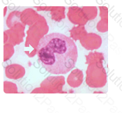
As maturation continues in the granulocytic series the nucleus of the metamyelocyte becomes kidney or bean shaped.
Identify the cell in this illustration indicated by the arrow:
Monoclonal antibodies are monospecific antibodies that are the same because they are made by one type of immune cell which are all clones of a unique parent cell, also called a hybrid cell line, which usually arise from a hybridoma. The fusion of a specific antibody-producing lymphocyte with a myeloma cell will multiply to become a source of pure monoclonal antibody. This is often used in the manufacturing process for monoclonal antibody reagents.
Monoclonal antibodies are usually manufactured in vitro by using:
If Kappa or Lambda is predominant and CD5 is co-expressed with CD19 (CD19/CD5 dual positive lymphocyte population), and CD23 is expressed, chronic lymphocytic leukemia is a probable diagnosis. CD19 is normally found on normal B cell populations and CD5 is normally found on mature T cell populations. However, CD5 is present on B cells in B-chronic lymphocytic leukemia or Mantle Cell Lymphoma, both abnormal B cell malignant processes.
CD23 expression is not consistent with Mantel cells as shown on the decision tree on the right.
A monoclonal B-cell population (Kappa or Lambda predominant) with expression of CD19, CD20, CD23, and co-expression of CD5 is consistent with which of the following?
Calcium oxalates can be found in normal urine in varying pH levels. Ammonium biurate and triple phosphate can be found in normal alkaline urine. Leucine crystals are always abnormal, and can be found in maple syrup urine disease.
Which one of the following crystals is NOT found in normal urine?